

AG Baltrusch IRTG

Imaging of collagen structure and its linkage sites to other proteins of the extracellular matrix by multimodal nonlinear microscopy
My research focuses on applying advanced optical imaging techniques, including multiphoton microscopy (MPM), fluorescence lifetime imaging microscopy (FLIM), and second-harmonic generation (SHG) imaging, to investigate biological processes in cells and tissues.
I combine these approaches to explore cellular metabolism, characterize ocular tissue features associated with aging and disease, and examine collagen organization and alterations in the extracellular matrix. My work integrates fluorescence lifetime measurements and phasor analysis with structural imaging to investigate both functional and morphological characteristics of biological samples.
Across these research areas, I am interested in how complementary optical measurements can reveal changes that are not readily apparent from conventional intensity-based images alone. The broader aim of my research is to connect cellular function with tissue structure and assess the potential of multimodal optical imaging for studying biological changes in health and disease.
| Autor | M. Sc. Samaneh Shahab |
|---|---|
| Betreuerin | Simone Baltrusch (UMR) |
| Zweitbetreuer | Stefan Lochbrunner (UR) |
| Kooperation | Barry Sanders (UCalgary, Kananda); Marcus Walckling (UMR); André Schulz (UMR); Mark Philipp (UMR) |
| Mehr erfahren | Samaneh Shahab - IRTG 2676 - Imaging Quantum Systems - Universität Rostock |
Quantifiable muscle imaging
Imaging is an important tool in muscle ageing research. Label-free approaches are particularly useful because they reduce sample preparation and allow tissue to be examined ex vivo without fluorescent staining. One such approach is multiphoton microscopy, which we use to image muscle tissue through its intrinsic two photon fluorescence and second-harmonic generation (SHG).
SHG arises from ordered tissue structures, including collagen and myosin. In muscle, the myosin signal reveals the repeating pattern of sarcomeres—the contractile units within muscle fibres. These images provide valuable visual insights, but quantitative measurements are needed to compare samples and assess whether observed differences are statistically significant.
My project takes two complementary approaches to this challenge. First, I am developing software to extract measurements from multiphoton images, including sarcomere length, muscle-fibre thickness and fibre orientation. Second, I use fluorescence lifetime imaging microscopy (FLIM) to measure how long naturally occurring fluorescent molecules emit light after excitation. These measurements may provide insights into metabolic changes associated with muscle ageing.
| Autor | M. Eng. Isabell Wenskus |
|---|---|
| Betreuerin | Simone Baltrusch (UMR) |
| Zweitbetreuer | Stefan Lochbrunner (UR) |
| Kooperation | Shabir Barzanjeh (UCalgary, Kanada); Oliver Friedrich (FAU, Erlangen); André Schulz (UMR) |
| Mehr erfahren | Isabell Wenskus - IRTG 2676 - Imaging Quantum Systems - Universität Rostock |
Understanding muscle function restriction with loss of mobility in old age
Aging is a universal process that affects everyone. In this context Sarcopenia plays an important role. It is characterized by an age-related loss of muscle strength and muscle mass, accompanied by a decline in physical performance. Since skeletal muscle plays a central role in maintaining physical performance and quality of life, age-related changes in the musculature can have far-reaching consequences.
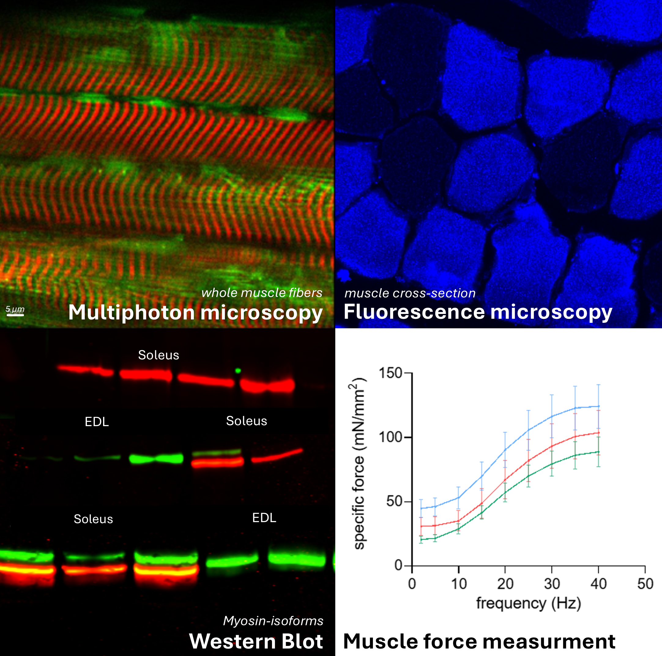
A deeper understanding of these underlying processes at a molecular level is intended to provide new insights into prevention and treatment of age-related diseases. To contribute to this understanding, I combine molecular biological methods with state-of-the-art microscopy techniques. In particular, I investigate the differences between the fatigue-resistant muscle soleus, a deep-seated calf muscle, and the more rapidly fatigued musculus extensor digitorum longus, which runs along the shinbone. The focus here is on the age-dependent changes in skeletal muscle, which are to be investigated muscle-specific and sex-specific.
| Autor | M. Sc. Chiara Buchsteiner |
|---|---|
| Betreuerin | Simone Baltrusch (UMR) |
| Zweitbetreuer | Stefan Lochbrunner (UR) |
| Kooperation | Pina Colarusso (UCalgary, Kanada); Gabriele Pfitzer (UKK, Köln) |
| Mehr erfahren | Chiara Buchsteiner - IRTG 2676 - Imaging Quantum Systems - Universität Rostock |